CT Scans and Cancer Risk: What the Evidence Shows
Key takeaways · 13 min read
- A CT scan gives far more radiation than an ordinary X-ray. For one person the added cancer risk is small, and most of what we know about it is estimated, not counted.
- A 2025 model projected about 103,000 lifetime cancers from the 93 million US CT scans of 2023. It relies on atomic-bomb survivor data and assumes no safe threshold.
- Studies of children scanned in Britain, Australia and Europe found more leukaemia and brain cancer at higher doses: roughly one or two extra cases per 10,000 children scanned.
- Part of that excess reflects why children were scanned: early tumour symptoms or conditions that raise cancer risk anyway. How large a part is still disputed.
In this article
- How much radiation a CT scan gives
- The projection of 103,000 cancers
- What the child cohort studies found
- Age, dose and the part of the body
- Reverse causation and confounding
- The linear no-threshold debate
- Overuse and the dose lottery
- The risk of not scanning
- What a scan costs in dose, and what to ask
- Questions people ask
- The short version
- Sources
In the 1980s American hospitals did about 3 million CT scans a year. In 2023 they did around 93 million. CT is now the default for a bad headache in the emergency department, a fall or a pain in the belly. It also uses X-rays, and X-rays at high enough doses cause cancer.
In April 2025 a team led by Rebecca Smith-Bindman at the University of California, San Francisco, put a number on the trade-off: about 103,000 future cancers from one year of American scanning. An earlier post covered thyroid cancer overdiagnosis, another side effect of the imaging boom.
The answer has two parts. The adult figures are projections from a model whose key assumption is sensible for radiation protection but unproven at these doses. The child figures come from real follow-up, but scans are not handed out at random. And a scan a doctor genuinely needs is almost always worth far more than its risk.
How much radiation a CT scan gives
A CT scanner builds cross-sections from hundreds of X-ray views, and that detail costs dose. The US Food and Drug Administration (FDA) gives typical effective doses of about 2 millisieverts (mSv) for a head CT, 7 for the chest and 8 for the abdomen, against 0.02 for a chest X-ray. Real doses can be two or three times higher or lower.
UW Medicine describes a chest CT as roughly two years of natural background radiation. On the conservative model used in radiation protection, the FDA says a 10 mSv scan might add one fatal cancer per 2,000 people. Of those 2,000, about 400 would die of cancer anyway.
Children get more dose per organ and have more years ahead of them. In the 2025 US analysis, a routine head CT gave boys under one an average brain dose of 60 milligray (mGy).
The projection of 103,000 cancers
Smith-Bindman’s team took scan-level data from 143 US hospitals and imaging centres, estimated organ doses, and scaled them to the 93 million scans of 2023 in about 62 million people, leaving out the 10.6% done in a patient’s last year of life. They then applied the National Cancer Institute’s risk tool, built on the BEIR VII models.
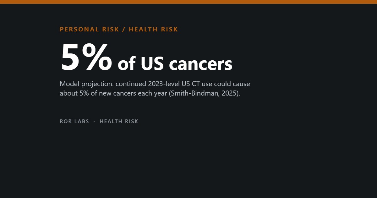
The result was 102,700 projected lifetime cancers (uncertainty range 96,400 to 109,500): 93,000 from adult scans and 9,700 from children. If scanning stayed at that level, CT could account for about 5% of new cancers each year. Other assumptions gave 80,000 to 127,000.
What the 2025 model projects
Projected lifetime cancers from one year of US CT scanning (2023), leading types for adult and child scans.
Source: Smith-Bindman et al., JAMA Internal Medicine (2025). Model projections using BEIR VII risk coefficients, not observed cases.
Per scan, babies carried the most risk: 20 cancers per 1,000 examinations in girls under one, against 2 per 1,000 in girls aged 15 to 17. Yet adults produced 90% of the total, because they have so many more scans. Abdomen and pelvis CT alone made up 40% of the adult projection.
This is modelling. BEIR VII rests mainly on survivors of the Hiroshima and Nagasaki bombs, exposed in a single flash; direct estimates for adult CT do not exist. The work was funded by the National Cancer Institute and the Patient-Centered Outcomes Research Institute. Smith-Bindman also co-founded a company that makes dose-monitoring software, as KFF Health News reported.
What the child cohort studies found
For children, researchers have counted rather than modelled. Mark Pearce’s 2012 Lancet study followed 178,604 people in Britain first scanned before the age of 22. Bone-marrow doses of 30 mGy or more went with about three times the leukaemia risk of doses under 5 mGy, and brain doses of 50 to 74 mGy with 2.82 times the brain-tumour risk. In absolute terms: one extra leukaemia and one extra brain tumour per 10,000 head scans in children under ten, over a decade.
In 2013 John Mathews and colleagues linked Medicare records for 10.9 million young Australians. The 680,211 scanned before 20 had 24% more cancers than those who were not, and the excess grew by 0.16 with each extra scan. That is about one excess cancer per 1,800 scans.
The European EPI-CT study pooled records from nine countries. In 2023 it reported an excess relative risk of 1.27 per 100 mGy for brain cancer in 658,752 young people, and 1.96 for blood cancers in 948,174. At today’s average doses, it expects one or two extra blood cancers per 10,000 children scanned over the next 12 years.
Different countries, similar slopes
Excess relative risk per 100 mGy of organ dose in cohort studies of children and young people scanned with CT. Higher means a steeper rise in risk with dose.
Sources: Pearce et al., Lancet (2012), per-mGy values x100; Smith-Bindman et al., NEJM (2025); Hauptmann et al., Lancet Oncology (2023); Bosch de Basea Gómez et al., Nature Medicine (2023); Meulepas et al., JNCI (2019). Confidence intervals are wide in all.
Age, dose and the part of the body
The pattern looks like radiation. In EPI-CT the relative risk of brain cancer was 2.1 at 41 to 47 mGy and 5.0 at 150 mGy or more, compared with under 5 mGy. In Australia the excess was largest in children first scanned before five, and cancers tended to follow the part scanned: leukaemia rose most after abdominal or pelvic scans, brain cancer after head scans.
The largest study, published in the New England Journal of Medicine in September 2025, followed 3.7 million children in six US health systems and Ontario. Blood-cancer risk rose with cumulative dose from all imaging, to a relative risk of 3.59 at 50 to 100 mGy, and the authors attributed 10.1% of the 2,961 blood cancers to imaging radiation.
Not every result lines up. In a Dutch cohort of 168,394 children, bone-marrow dose was not linked to leukaemia at all, although brain dose was linked to brain tumours. And in Australia more than half the excess cancers occurred in people first scanned at 15 to 19, because nearly half of those scanned were teenagers.
Reverse causation and confounding
Scans are not given at random. A child with headaches from a slow-growing brain tumour may have a head CT years before the diagnosis; then the cancer caused the scan, not the other way round. Children with conditions such as Down syndrome or tuberous sclerosis are scanned more often and develop some cancers more often, for reasons unrelated to X-rays.
Researchers handle this by ignoring the first years after a scan. In Australia, stretching that gap from one year to ten shrank the excess from 24% to 18%, and the count of excess cancers from 608 to 209. Brain cancer gave the clearest warning sign: its excess was largest one to four years after the first scan, then faded.
A 2013 report by the UN Scientific Committee on the Effects of Atomic Radiation (UNSCEAR) concluded that the associations in the early studies may have come from confounding by indication rather than radiation, as John Boice summarised in 2015.
Yet brain cancers after scans of other body parts were still raised in Australia, and removing children with tuberous sclerosis did not change the Dutch brain-tumour result. A 2016 modelling study judged that cancer-prone syndromes do not meaningfully distort leukaemia risks, though one of them could inflate brain-tumour risks.
The excess shrinks with time, but does not vanish
Australian cohort: incidence rate ratio for cancer in people scanned before age 20 compared with people not scanned. 1 = no difference.
Source: Mathews et al., BMJ (2013); 680,211 exposed people among 10.9 million. Ratios compare groups; they are not absolute risks.
The linear no-threshold debate
The linear no-threshold model assumes that cancer risk rises in a straight line with dose, all the way down to zero. Radiation protection has used it since the late 1950s. The International Commission on Radiological Protection calls it a prudent basis for low doses, and in 2021 the US Nuclear Regulatory Commission rejected petitions to drop it.
There is evidence behind it. DNA double-strand breaks rise in a straight line with dose down to about 10 mGy, and Mathews’s per-dose risks for leukaemia and brain cancer were compatible with the bomb-survivor estimates. A 2023 review from the French institute IRSN found that a decade of new data had reinforced the evidence of risk at 100 mGy or less, though some cancers may not follow a straight line.
The critics are not fringe. A Society of Nuclear Medicine and Molecular Imaging task group concluded in 2019 that the model is not supported at low doses, and six of seven animal studies reviewed by UNSCEAR in 2021 showed no rise in tumours at their lowest dose. Nobody can measure the risk of a single scan directly.
Overuse and the dose lottery
Whatever the exact risk, unneeded scans and needlessly high doses add to it with no benefit. A 2019 BMJ study of more than 2 million adult scans at 151 institutions in seven countries found that average doses for the same scan varied several-fold, mainly because of how local staff set the machines rather than differences between patients.
Same scan, different dose
Lowest and highest country averages of effective dose for adult CT, across seven countries.
Source: Smith-Bindman et al., BMJ (2019); more than 2.0 million examinations, 151 institutions, seven countries, 2015–2017.
Children are not always better protected. A 2025 US study of 192,185 scans found teenagers’ head and abdominal doses 20 to 30% lower in children’s hospitals, but chest doses for under-12s 30 to 100% higher. In the 2025 projection, 28.5% of examinations were multiphase, scanning the same area more than once, which often could be avoided.
Some scans are simply not needed. The American Academy of Pediatrics says about half of children seen in emergency departments with a head injury get a CT, many unnecessarily, and recommends a period of observation first. In 2025 Medicare proposed keeping its CT dose quality measure for hospitals voluntary. Doses are falling anyway, as the news above shows.
The risk of not scanning
Every number above concerns the scan. None of them counts what happens without it: a bleed in the brain found late, an appendix that bursts, a cancer staged too late. The FDA lists the small cancer risk among the downsides of CT, alongside incidental findings that lead to more tests, but calls it much smaller than a person’s natural risk of cancer.
Lung screening shows the balance. In the US National Lung Screening Trial of 53,454 heavy smokers, low-dose CT cut lung cancer deaths by 20% compared with chest X-rays. A Dutch model of women who smoked heavily and were screened every year from 55 to 75 estimated 112.4 cancers found per 1,000, against 2.2 caused by the radiation.
Benefit and risk on the same scale
Two published comparisons. The radiation figures come from linear no-threshold models.
Sources: Du et al., European Journal of Radiology (2022), modelling of heavy smokers; US Food and Drug Administration, ‘What are the radiation risks from CT?’
Pearce’s team wrote that the clinical benefit should outweigh the small absolute risk, as long as doses are kept as low as possible. The worry is the scan ordered just to see, not the one that answers a real question. If a doctor recommends CT, ask why. Do not refuse it out of fear.
What a scan costs in dose, and what to ask
Doses are measured in millisieverts (mSv). The US Food and Drug Administration (FDA) lists these average effective doses.
| Exam | Dose (mSv) |
|---|---|
| Chest X-ray | 0.02 |
| CT head | 2 |
| CT coronary calcium | 3 |
| CT chest | 7 |
| CT abdomen | 8 |
| CT coronary angiogram | 16 |
How big is the risk? The FDA says a 10 mSv CT may raise the chance of a fatal cancer by about 1 in 2,000, using a linear no-threshold model, against a natural fatal cancer risk of about 1 in 5. It calls the risk very small but statistically uncertain. The National Cancer Institute (NCI) compares a low-dose chest CT (1.5 mSv) with about six months of natural background radiation and a typical chest CT (6.1 mSv) with about two years.
Who it applies to most. Children are more sensitive; the NCI cites an excess risk of about 1 to 2 cases per 10,000 scans over 12 years after a single CT. Women are at somewhat higher risk than men after the same exposure at the same age. The NCI says CT radiation is believed to be too low to harm a growing fetus.
What to ask. The NCI suggests parents ask why the test is needed, whether the results will change treatment, and whether an alternative without radiation exists. The FDA says to discuss any earlier X-ray procedures, lists ultrasound and MRI as lower-radiation options, and sees no benefit in whole-body scanning of people without symptoms.
What we could not confirm. The FDA risk page dates from 2017 and its 1-in-2,000 figure is not repeated by the NCI, whose 2024 fact sheet gives no lifetime-risk number, so the estimates differ by source. The FDA main page links out for pregnancy advice rather than stating it.
What to do
Steps sorted by when they matter. Each one says how strong the evidence is: Measured Official advice Untested
Before
- Ask your doctor whether the scan is needed and what it is expected to show. Untested see Sources
- Ask whether an ultrasound or MRI, which use no radiation, could answer the same question. Untested see Sources
- Tell the doctor about CT scans you or your child have had before, using a written imaging record. Official advice see Sources
- For a child with a minor head injury, ask whether a period of observation could come before a CT. Official advice see Sources
If it happens
- For a child, ask whether the scan settings are adjusted for the child’s size. Untested see Sources
- Ask whether the area needs to be scanned more than once; many multiphase scans could be done in a single pass. Measured see Sources
Afterwards
- Add the scan, its date and where it was done to your or your child’s imaging record. Official advice see Sources
- Share that record with future doctors so they can weigh it before ordering another scan. Official advice see Sources
- Ask what any incidental finding means before agreeing to more tests, since such findings often lead to further testing. Official advice see Sources
What not to do
- Do not refuse or delay a CT scan your doctor recommends, especially in an emergency. Untested see Sources
- Do not read the 103,000 figure as a count of real cases; it is a model projection. Measured see Sources
- Do not ask for extra scans just for reassurance; a scan that changes nothing adds risk without benefit. Untested see Sources
Not medical advice. Never refuse a scan your doctor recommends; ask whether it is needed.
Questions people ask
I have had several CT scans. Should I worry?
For most people, no. The risk from each scan is small. Keep a list of the scans you have had, so the next doctor can take it into account.
Is a CT scan more risky for my child?
Per scan, yes: children are more sensitive to radiation and have more years ahead. The child studies suggest one or two extra cancers per 10,000 children scanned. Ask whether ultrasound or MRI could answer the question, and whether the dose is set for a child’s size.
Do MRI and ultrasound carry the same risk?
No. Neither uses ionising radiation. They are not always the right test, but for suspected appendicitis in a child, Smith-Bindman notes, ultrasound is often a very good one.
Is 103,000 cancers a real count?
No. It is a projection from a risk model applied to one year of scans. The true figure could be lower or higher.
Should I refuse a CT scan in an emergency?
No. In an emergency, missing a bleed, a clot or a ruptured organ is an immediate danger, far larger than a small lifetime cancer risk. Ask what the scan is for, but do not delay it.
The short version
- A CT scan gives far more radiation than an ordinary X-ray. For one person the added cancer risk is small, and most of what we know about it is estimated, not counted.
- A 2025 model projected about 103,000 lifetime cancers from the 93 million US CT scans of 2023. It relies on atomic-bomb survivor data and assumes no safe threshold.
- Studies of children scanned in Britain, Australia and Europe found more leukaemia and brain cancer at higher doses: roughly one or two extra cases per 10,000 children scanned.
- Part of that excess reflects why children were scanned: early tumour symptoms or conditions that raise cancer risk anyway. How large a part is still disputed.
- Doses for the same scan vary several-fold between hospitals and countries, mostly because of machine settings. Unneeded scans and excess dose are the avoidable part of the risk.
- When a doctor recommends CT for a real clinical question, the benefit almost always outweighs the risk. Ask whether it is needed or whether a non-radiation test would do; never refuse out of fear.
This article summarises research, agency guidance and news reports for general information. It is not medical advice. Never refuse or delay a scan your doctor recommends because of this article; if you have questions about a planned scan, ask your doctor or the radiology team.
Further reading: Smith-Bindman et al., JAMA Internal Medicine (2025), for the US projection and its assumptions. Mathews et al., BMJ (2013), for the clearest look at reverse causation in the child data. Laurier et al., Journal of Radiological Protection (2023), for a balanced review of the linear no-threshold debate.
Sources
- Smith-Bindman R, et al. Projected lifetime cancer risks from current computed tomography imaging. JAMA Intern Med 185(6) (2025), published online 14 April 2025.
- Richman IB, Katz MH. Balancing computed tomography’s benefits with radiation risks (editor’s note). JAMA Intern Med 185(6) (2025).
- Pearce MS, et al. Radiation exposure from CT scans in childhood and subsequent risk of leukaemia and brain tumours: a retrospective cohort study. Lancet 380(9840):499–505 (2012), doi:10.1016/S0140-6736(12)60815-0.
- Mathews JD, et al. Cancer risk in 680 000 people exposed to computed tomography scans in childhood or adolescence: data linkage study of 11 million Australians. BMJ 346:f2360 (2013).
- Hauptmann M, et al. Brain cancer after radiation exposure from CT examinations of children and young adults: results from the EPI-CT cohort study. Lancet Oncol 24:45–53 (2023), doi:10.1016/S1470-2045(22)00655-6.
- Bosch de Basea Gómez M, et al. Risk of hematological malignancies from CT radiation exposure in children, adolescents and young adults. Nat Med (2023), doi:10.1038/s41591-023-02620-0.
- Smith-Bindman R, et al. Medical imaging and pediatric and adolescent hematologic cancer risk. N Engl J Med (published online 17 September 2025), PMID 40961449.
- Meulepas JM, et al. Radiation exposure from pediatric CT scans and subsequent cancer risk in the Netherlands. J Natl Cancer Inst (2019), doi:10.1093/jnci/djy104.
- Meulepas JM, et al. Confounding of the association between radiation exposure from CT scans and risk of leukemia and brain tumors by cancer susceptibility syndromes. J Radiol Prot 36(4):953 (2016), doi:10.1088/0952-4746/36/4/953.
- Boice JD Jr. Radiation epidemiology and recent paediatric computed tomography studies. Ann ICRP (2015), doi:10.1177/0146645315575877.
- Laurier D, et al. The scientific basis for the use of the linear no-threshold (LNT) model at low doses and dose rates in radiological protection. J Radiol Prot (2023), doi:10.1088/1361-6498/acdfd7.
- Siegel JA, et al. A critical assessment of the linear no-threshold hypothesis: its validity and applicability for use in risk assessment and radiation protection. Clin Nucl Med (2019), doi:10.1097/RLU.0000000000002613.
- Smith-Bindman R, et al. International variation in radiation dose for computed tomography examinations: prospective cohort study. BMJ 364:k4931 (2019).
- Mahendra M, et al. Cancer risk associated with radiation doses used for CT scans in pediatric and general hospitals. Hosp Pediatr (2025), doi:10.1542/hpeds.2024-008256.
- Du Y, et al. Lung cancer screening with low-dose CT: simulating the effect of starting screening at a younger age in women. Eur J Radiol (2022), doi:10.1016/j.ejrad.2022.110182.
- Aberle DR, et al. Computed tomography screening for lung cancer: has it finally arrived? Implications of the National Lung Screening Trial. J Clin Oncol (2013), doi:10.1200/JCO.2012.43.3110.
- US Food and Drug Administration. What are the radiation risks from CT? (web page, content current as of 5 December 2017).
- American Academy of Pediatrics. Choosing Wisely recommendation on CT for minor head injuries in children, as summarised in American Family Physician, Choosing Wisely collection.
- Image Gently. My Child’s Medical Imaging Record (parent materials, reproduced by NC Department of Health and Human Services).
- Kenen J. Some CT scans deliver too much radiation, researchers say. Regulators want to know more. KFF Health News (12 March 2025).
- Applied Radiology. CMS proposes ongoing voluntary status for CT radiation dose quality measure amid physician concerns (12 September 2025).
- Madden Yee K. Smith-Bindman unpacks NEJM pediatric CT study. AuntMinnie (25 September 2025).
- UW Medicine Newsroom. CT scan radiation doses fell substantially in past decade (18 August 2026).
- FDA. What are the radiation risks from CT? fda.gov.
- FDA. Computed tomography (CT). fda.gov.
- NCI. Computed tomography (CT) scans and cancer, reviewed 8 February 2024. cancer.gov.